Introduction
Nanotechnology is great because of all its practical applications, but what would really help scientists now (especially for biologists), would be to see nanoscale particles in order to observe an objects reactions. Even though scientists have been able to improve resolution through fluorescence microscopes, scientists are still not satisfied.
However there is a problem with traditional microscopes. they use light to make the object being viewed bigger by increasing its resolution. Basically the resolution is what comes up on the screen. if you zoom to much, the image is blurry because there is not enough of the little squares in the image to fit into the screen. However, if the little squares that make up that image are smaller, then you can zoom in more without the image getting blurry.
But, there is a problem. Light travels with a wave length of about 400nm. Scientists want a better resolution than that, and there in-lies the obvious conundrum. How are you supposed to see something that is smaller than light? at that size, two of the waves would overlap each other.
Fluorescence Microscopy

This is a broad range of scopes that use fluorescent light. The one above is the simple and original type - the Wide field microscope. Until recently, this has been the best choice for microscopy, though it has been brilliantly out shined since. It was quite simple. The fluorescent light excites all the particles of dye in the sample specimen, illuminating it all at once. Then the light is picked up by a camera or what is being used to see the specimen.
Now
The ones that we will discuss are the ones that "have high potential for cellular imaging and are commercially available" The Annual Review of Cell and Developmental Biology pg. 285 and still use light.
These are:
- Wide-Field confocal
- TIRF
- Structured Illumination
- STED
- Pointillism
Wide-Field Confocal Microscope

Wide-Field confocal was the big breakthrough after wide field microscopes. This type of microscope emits lasers or Fluorescent light to take point size images and then putting it together like a scanner, unlike the Fluorescence Microscopes which take the pictures like a camera. This leads to the limitation of how small an area one can excite at a time. the smaller of an area that is excited, the better resolution there is. Some Wide-Field Confocal Microscopes even have multiple lasers to color different parts of a cell.
Because it uses lasers, it can actually go through layers of a cell meaning that by doing many pictures with slightly different z-plane dimensions, computers can create clear 3D images with it.
However, it is not the best microscope out there. Wide-Field confocals have a limit of 200nm resolution (about 1/2 the resolution of light.)
TIRF
TIRF to be continued
STED
Stimulated emission depletion microscopy (STED) is one of the best types of microscopes there is. It can reach a resolution of 5.8nm
To be continued
Pointillism
Is basically the best way we have to go nanoscale with light. When objects get smaller than the wave length of light, two waves will overlap on it. Basically, a computer will then transform the image into an equation, then create another equation to put the overlap together into a clear image.
Although this is the best way to get maximal resolution, it bleaches, overall, is not good for seeing animals, whereas the wide-field Confocal is.
and my dad (Thank you)
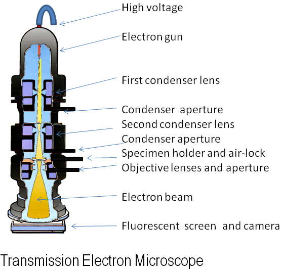
Electron Microscope
This method does not use light, but electrons.

Nano is 10 to the -9th meters. Nanoscale is anything from 1-100 nanometers. Atoms are roughly 10 to the -10 meters. That means that 10 atoms fit in one nanometer and 2,000 atoms is the smallest resolution of a wide-Field Confocal. In short, it is tiny, and no light-beam-based scope can see it. However, we have discovered something else. Where as the microscopes above use light, or beams of photons, electron microscopes use beams of electrons. To get beams of electrons, individual electrons need to be isolated. To do this, a piece of Tungsten is heated up. As learned in basic physics, atoms heat up because the electrons are speeding up and getting excited, and when the electrons get excited, they can move out of their ring (Bohr Model). At this point the atom is known as an ion because the number of atoms and electron are not equal.
 |
| A tungsten atom. when excited, it will lose an atom which will then get shot out by the electron gun. |
An electron gun is two electrodes, that are close to each other, and create and electric field. This field accelerates the electrons creating a beam. This beam is then shot into a vacuum. This vacuum is quite important in this microscope because if it weren't there the electrons would scatter in the same way the alpha particles did in Rutherford's famous gold foil experiment. To learn more, click here.
 |
| Rutherford's famous gold foil experiment |

To achieve even more efficiency, there are a series of 'condenser lenses', more formally known as electromagnetic lenses, which focus the beam of electrons even further by changing the direction of the beam.

Once there is an electron beam going and focused, the specimen being observed must be coated with a metallic substance in order for the electrons to bounce off, on to a screen (phosphorescent screen, layer of photographic film, or sensor such as a CCD camera) where the image is then displayed.
Unfortunately, it can only display surface images, though it can eventually produce 3D in addition to 2D images. It is also becoming possible to add color.
http://www.microscopyu.com/articles/fluorescence/tirf/tirfintro.html\http://serc.carleton.edu/microbelife/research_methods/microscopy/fluromic.html